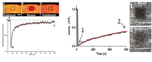
Confocal methods can also be used to measure the mobility of fluorescent molecules in the cytoplasm of muscle cells, in a process called “FRAP” (short for “fluorescence recovery after photobleaching”). In FRAP, a molecule or complex is tagged with a fluorescent marker and then a region that contains the marker is bleached with a brief pulse of intense laser light. The bleached region is then followed under confocal optics over time to measure the rate and extent to which the fluorescence signal recovers. Two parameters can be determined from this experiment: the % of the originally bleached molecule or complex that is mobile, and the half life of recovery of the mobile fraction, which is related to the diffusion coefficient, D. When we used FRAP to measure the mobility of the Fluo5N-Ca2+ complex in muscle fibers, we found the mobile fraction to be ~100% and diffusion to occur with a half life of recovery of ~3 sec (see left panel below). By contrast, when we used FRAP to measure the mobility of a fluorescent chimera of dysferlin in muscle fibers, the mobile fraction was only ~40%, with half life of recovery of ~330 sec (see right panels below). We plan to use FRAP to study the characteristics of the proteins of the SR and the t-tubules and to investigate the patterns by which they travel through the cytoplasm to reach their final destinations.
We are currently experimenting with mobility studies that afford resolution at the level of single molecules, with a method called PALM (photoactivation light microscopy). In PALM, individual fluorescent molecules are activated stochastically, by very low levels of confocal laser illumination, and then followed over time until the fluorescence decays. This method has the potential to reveal the patterns of trafficking of proteins of the SR and t-tubules in molecular detail.